



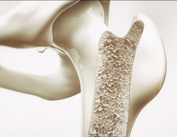
A new study generated one of the most comprehensive spatially resolved transcriptomic maps of cellular communication between bone and skeletal muscle in a young mouse. Using spatial transcriptomics, computational deconvolution, and ligand-receptor network analysis, researchers identified signaling pathways that coordinate tissue maintenance, remodeling, and vascular support. Key interactions involving collagen, thrombospondin, tenascin, and VEGF pathways were validated experimentally and across independent datasets. The findings provide a foundation for studying musculoskeletal disorders and aging.
Bone and skeletal muscle are often viewed as separate tissues with distinct functions, yet they operate as a highly integrated system. Together, they support movement, maintain posture, regulate metabolism, and help preserve overall health. Scientists have long known that bone and muscle communicate through biochemical signals, but understanding exactly where these molecular conversations occur and which cells participate has remained a major challenge. Traditional genomic technologies can identify genes expressed within tissues but often lose the spatial information needed to understand how neighboring cells interact within their natural environment.
Addressing this challenge, a research team was led by Professor Hong-Wen Deng, Director at the Tulane Center for Biomedical Informatics and Genomics, Deming Department of Medicine, School of Medicine, Tulane University, USA. The researchers applied spatial transcriptomics, an emerging technology that maps gene activity directly within intact tissues, to examine a mouse femur and its adjacent skeletal muscle. By combining this approach with advanced computational tools, they reconstructed cellular neighborhoods and communication networks across the bone-muscle interface. The analysis generated data from 2,660 spatial spots and identified multiple major cell populations participating in tissue communication. Their findings were published in Volume 14 of the journal Bone Research on May 19, 2026.
The study revealed that bone and muscle are linked through a surprisingly complex communication system involving osteoblasts, skeletal muscle cells, endothelial cells, immune cells, and stem-cell populations. Researchers identified thirteen major signaling pathways that coordinate tissue maintenance and remodeling. Several of these pathways involved extracellular matrix proteins and growth factors that help cells exchange information, regulate structural integrity, and respond to physiological demands. The findings suggest that communication between tissues is not random but organized into distinct spatial networks shaped by local cellular environments.
One of the study’s most significant discoveries was the identification of specific ligand-receptor pairs that act as molecular messengers between neighboring cells. These included collagen-associated signaling between osteoblasts and muscle cells, thrombospondin-mediated communication involving immune cells, and vascular endothelial growth factor (VEGF)-driven signaling that supports vascular function. Laboratory imaging confirmed the colocalization of several predicted molecular partners within the tissue, strengthening confidence in the computational predictions. Additional validation using independent mouse and human datasets supported many of the identified pathways, suggesting that some communication mechanisms may be shared across species.
“Our goal was to move beyond simply identifying which genes are present and instead understand how cells communicate within their native tissue environment,” explained Prof. Deng. “By preserving spatial information, we were able to uncover communication networks that would be difficult to detect using conventional sequencing approaches alone.”
The work also offers important opportunities for future collaboration across fields including bone biology, muscle physiology, regenerative medicine, aging research, bioinformatics, and precision medicine. Because disorders, such as osteoporosis, sarcopenia, and metabolic disease, often involve simultaneous deterioration of bone and muscle, a clearer understanding of tissue crosstalk could help researchers identify shared therapeutic targets. In the short term, the study provides a valuable reference map that scientists can use to investigate how these signaling networks change during injury, aging, or disease progression.
“Understanding these cellular communication pathways gives us a framework for studying what goes wrong in musculoskeletal disorders,” said Prof. Deng. “In the future, this knowledge may help guide the development of targeted interventions that restore healthy communication between tissues.”
Overall, the study delivers one of the first spatially resolved, transcriptome-wide maps of bone-muscle communication. By revealing how cells coordinate their activities through organized signaling networks, the research establishes a foundation for future investigations into musculoskeletal health and disease. Over the longer term, such insights could contribute to more precise diagnostic tools, improved regenerative therapies, and personalized treatment strategies aimed at preserving mobility and quality of life in aging populations.
Disclaimer: This content has not been generated, created or edited by Finance SC. Publisher:
Source link
Source link